1.Field-emission scanning electron microscope, SEM
Instruments
高解析場發射掃描式電子顯微鏡
|
The JSM-6500F, a field-emission scanning electron microscope, which employs a Schottky type field-emission (T-FE) gun for the electron source and state-of-the-art computer technology for the image-display system, has analytical functions such as EDX and CL, as well as a function for high-resolution image observation. The probe-current range is from several pA to 100 nA. The accerlated voltage is from 0.5 kV to 30 kV, with 1.5 nm (15 kV) spatial resolution. (From JEOL) |
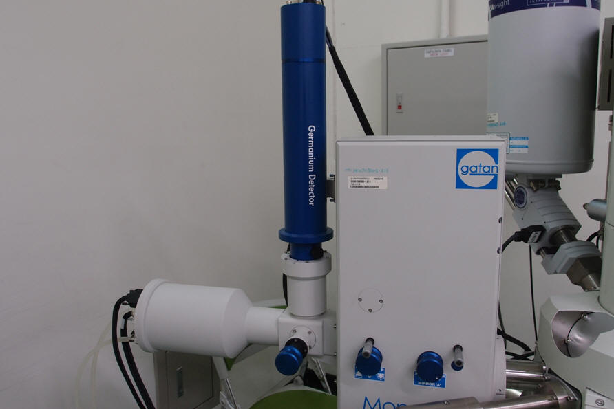 2.Cathodoluminescence system, CL
陰極螢光光譜及影像系統
|
|
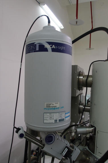 3.Energy dispersive
X-ray spectroscopy, EDX
3.Energy dispersive
X-ray spectroscopy, EDX
能量分散元素分析光譜儀
|
|
 4.Raman-scattering
spectroscopy
4.Raman-scattering
spectroscopy
拉曼光譜儀
|
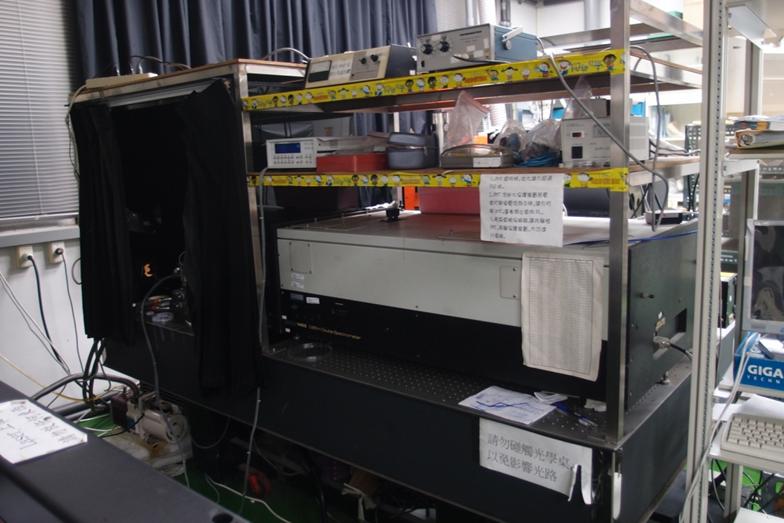
Raman spectroscopy is a spectroscopic technique based on inelastic scattering of monochromatic light, usually from a laser source. Inelastic scattering means that the frequency of photons in monochromatic light changes upon interaction with a sample. Photons of the laser light are absorbed by the sample and then reemitted. Frequency of the reemitted photons is shifted up or down in comparison with original monochromatic frequency, which is called the Raman effect. This shift provides information about vibrational, rotational and other low frequency transitions in molecules. Raman spectroscopy can be used to study solid, liquid and gaseous samples. |
5.Spectrophotometer
分光光譜儀
|
•Measurement
range: 190 nm – 3300 nm
•UV/Vis
Resolution: 0.17 – 5.00 nm
•NIR
Resolution: 0.20 – 20.00 nm
•With
a 60 mm integrating sphere
|
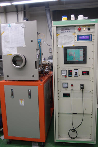
6.Thermal evaporator
熱蒸鍍機
|
|
 7.Atomic
Force Microscopy
7.Atomic
Force Microscopy
原子力顯微鏡
|
Atomic force microscope (AFM) is a type of scanning probe microscopes (SPM). It is operated by measuring force between a probe and the specimen surfaces. In general, the probe is a sharp tip at a cantilever's end. The cantilever can be deflected by atomic forces to sufficiently large amount, then AFM can measure the vertical and lateral deflections of the cantilever by using the optical system. A laser beam is transmitted to cantilever, and the reflected laser beam is detected with a position-sensitive photo detector (PSPD). The output of the PSPD is provided to a computer for processing of the data for providing a topographical image of the surface with atomic resolution, and controlling the height between probe and specimen surfaces by applying voltage on piezoelectric scanner.
|
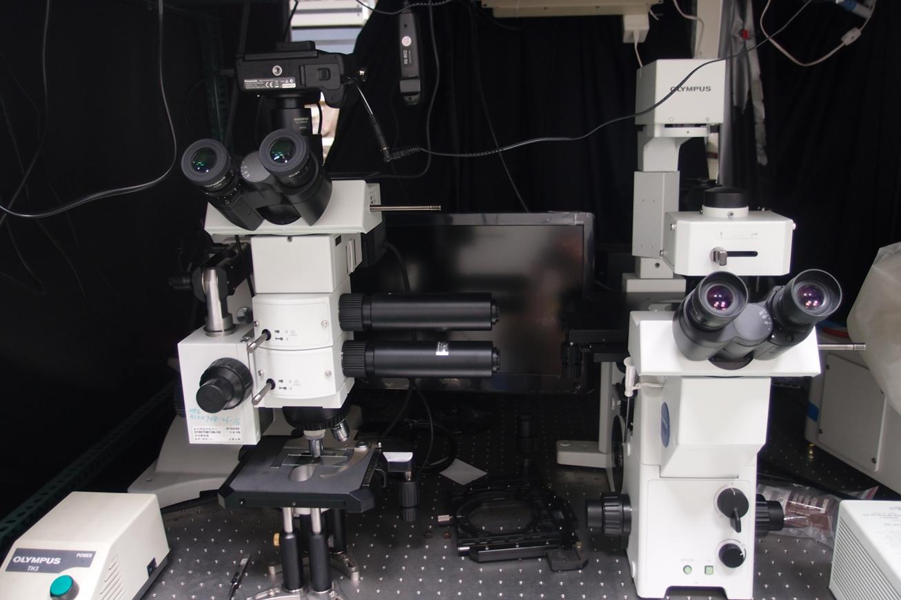
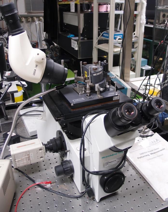
8.Optical Microscopy
光學顯微鏡
|
|
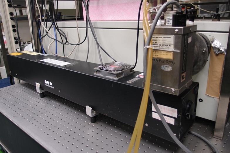 9.Photoluminescence
(PL) Measured System
9.Photoluminescence
(PL) Measured System
光激螢光量測系統
(325 nm He-Ne Laser上圖、Grating
system下圖)
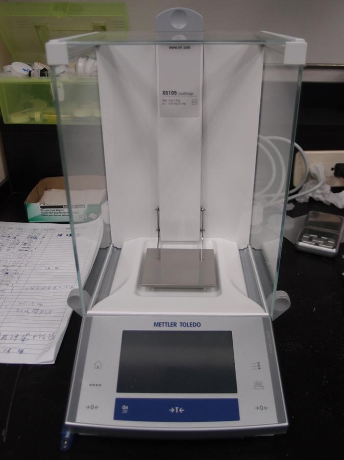
10.Electronic Balance
電子天平
|
|
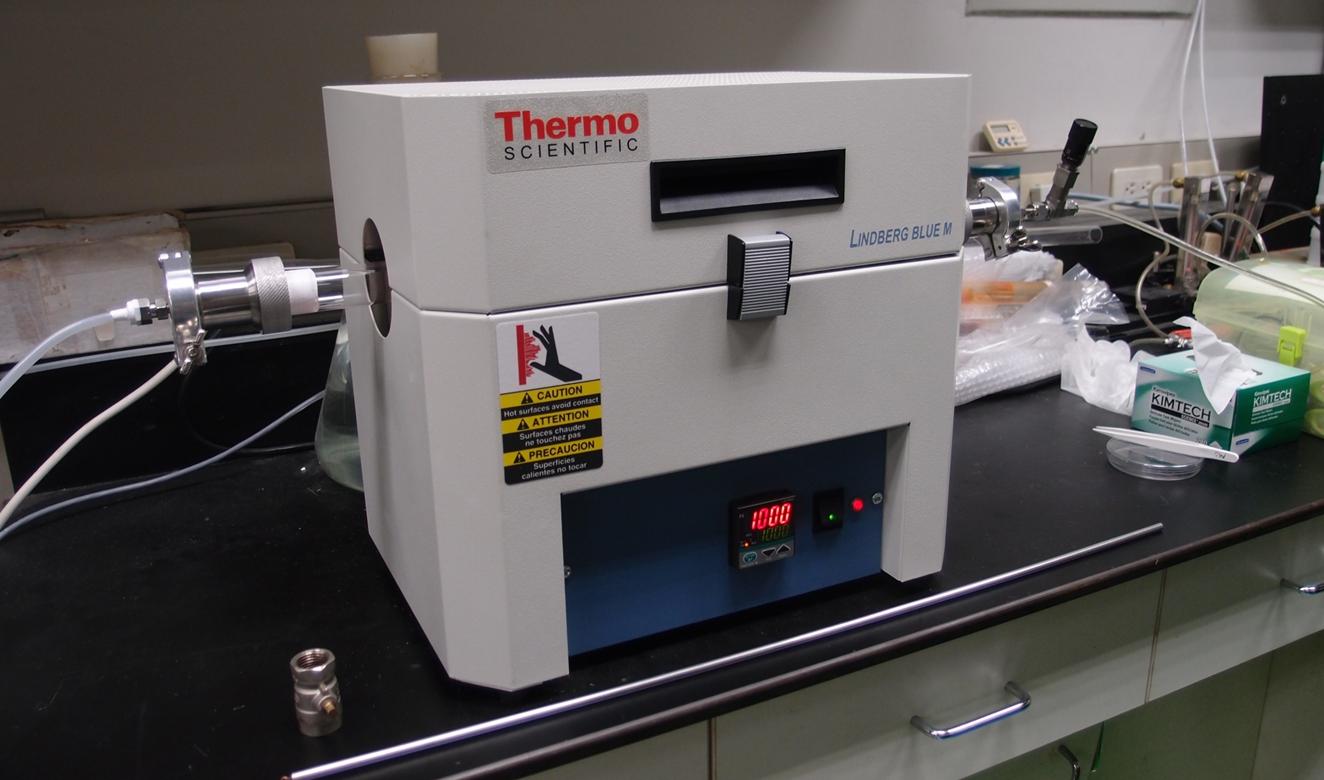 11.High
Temperature Furnace
11.High
Temperature Furnace
高溫爐
|
|
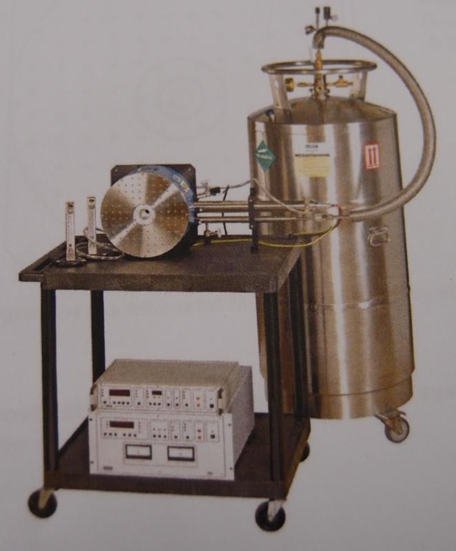 12.Microstat
Magneto-optical (MO) System
12.Microstat
Magneto-optical (MO) System
高磁場微觀光學量測系統
|
The Microstat MO is a continuous flow liquid helium cryostat combined with a conduction cooled superconducting magnet. The design allows a sample to be cooled to a low temperature and studied in a magnetic field both optically and electrically simultaneously. The window arrangement allows the sample to be brought close to the objective lens of the microscope with the sample mounted in vacuum and cooled by conduction. A second window in the base of the cryostat enables the possibility of transmission measurements to be performed. (From Oxford Instruments)
|
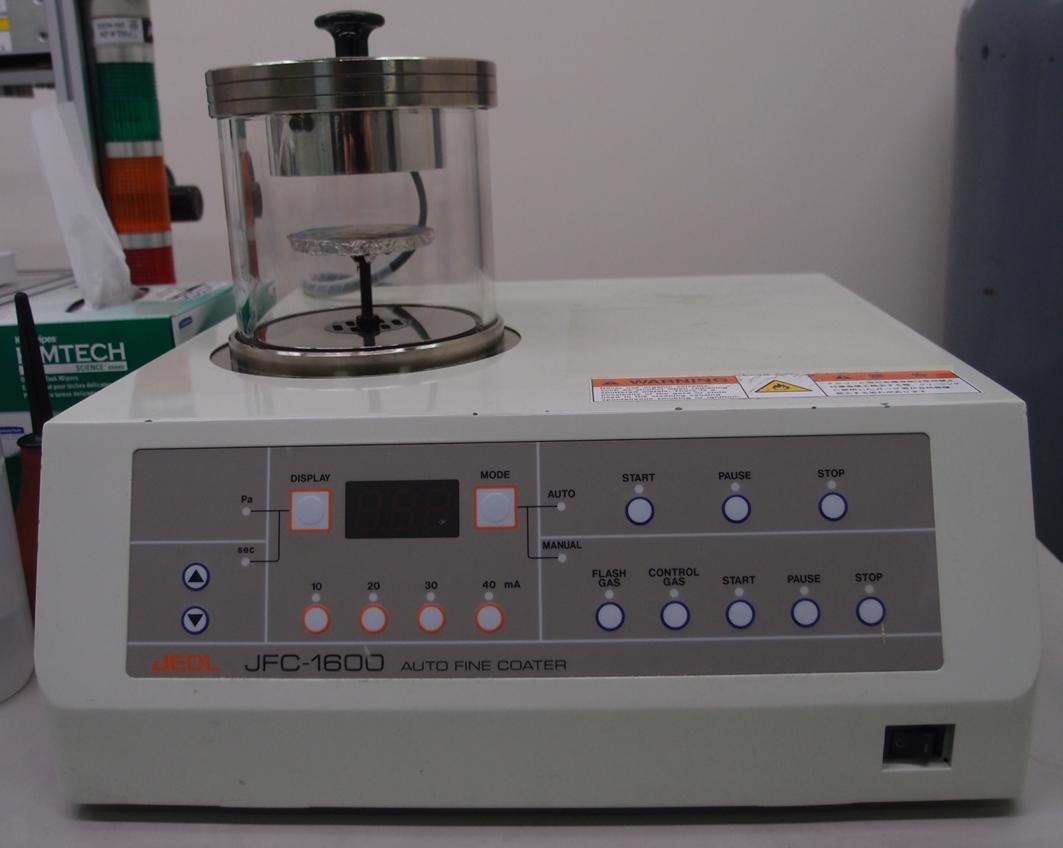 13.Auto
Fine Coater
13.Auto
Fine Coater
鍍金機
|
This coater, which consists for a main unit and a pump, is intended
mainly to preparing specimens for SEM observation.
|